Written By: Jeff Rogge, PT, DPT, OCS, CMTPT, FAAOMPT
Hip pain caused by femoroacetabular impingement (FAI) can be very painful and frustrating for our patients. FAI occurs when there is bony overgrowth around the femoral head (Cam) and/or around the acetabulum (Pincer.) Currently, we are not able to predict which individuals with Cam or Pincer morphologies will develop FAI syndrome.
When it comes to FAI it is important to view it as a Syndrome. FAI is considered a normal variation of hip morphology and is often present on imaging in asymptomatic patients. We cannot predict who will have pain or issues in the future strictly based on the findings on imaging. To accurately diagnose FAI it is important to triangulate the findings of imaging, testing, and the patient’s symptoms.
The idea in physical therapy known as regional interdependence is the concept that seemingly unrelated impairments in a remote anatomical region may contribute to, or be associated with, the patient’s primary complaint. I.e., TLJ syndrome and DF limitations are important to consider.31 With physical therapy treatment for hip FAI it is important to remember that similar to osteoarthritis we cannot undo the bony abnormalities. It is important to evaluate the patient’s neuromuscular control and motor planning of the hip complex.
When treating a patient with FAI it is important to access their acetabular plane. This is the same idea as scaption in the shoulder. The acetabular plane is the most stable position of the hip. The acetabular plane is stronger and more balanced with internal and external rotation strength in comparison with standard strength testing in which internal rotation is often substantially weaker. The patient will also have improved neuromuscular control in the scapular plane. The scapular plane can have significant variations from patient to patient.1
 In contrast, if a patient is demonstrating movements not in the acetabular plane, they will often have decreased strength, decreased neuromuscular control, decreased ROM, and increased potential for injury such as FAI, SIJ dysfunction, LBP, etc.
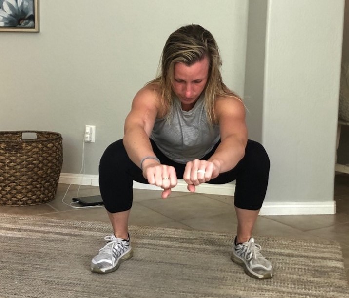
Squatting mechanics is important in patients with FAI. A study by Fry et al limited forward knee travel during a squat, or the typical knee behind your toes and feet pointed forward squat.2 This simply shifted the stress from the knees to the hips/low back. So, while squatting with restricted forward knee movement showed a 22% decrease in knee torque, there was a 1070% increase in hip torque! This is a LOT more work for the hip and low back musculature to perform.
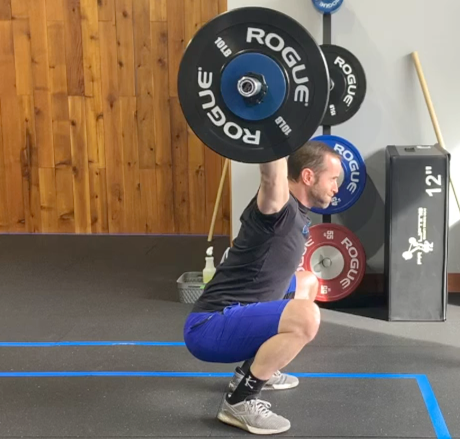 Patients with FAI have also been shown to often squat with increased lordosis creating early hip end range flexion
When considering treatment options for patients with FAI along with typical treatments of assessing pelvic floor, core stability, ROM, and strength deficits it is vital to improving body mechanics and neuromuscular control during strengthening and functional activities. It is important to focus on movement coordination deficits and increase neuromuscular control and motor planning of the hip. The stability of the pelvis on the femur demands a high degree of pelvic motor control. Addressing posterior hip soft tissue and joint mobility to decrease stress on the anterior hip is also an important consideration.
Be sure to register for my upcoming Live Webinar, Examining Evidence-Based Treatment Options for Common Hip Pathology on March 30th. This course will take you through the associated patient characteristics, evaluation findings, and radiological criteria needed for an accurate diagnosis. You will leave this course with a full understanding of how this condition is best managed as a syndrome.
Explore continuing education courses from Jeff below:
Advanced Concepts and Application for Manual Therapy of the Spine
Advanced Management of the Hip Joint Complex
Advanced Management of the Shoulder Girdle Complex
Examining Evidence-Based Treatment Options for Common Hip Pathology
Visit summit-education.com for more information.
References:
Fry, A.C., J.C. Smith, and B.K. Schilling, Effect of knee position on hip and knee torques during the barbell squat. J Strength Cond Res, 2003. 17(4): p. 629-33.
Thelen, T., et al., Normative 3D acetabular orientation measurements by the low-dose EOS imaging system in 102 asymptomatic subjects in standing position: Analyses by side, gender, pelvic incidence and reproducibility. Orthop Traumatol Surg Res, 2017. 103(2): p. 209-215.